
Foot injuries, whether from sports or accidents, are common, and MRI (Magnetic Resonance Imaging) plays a crucial role in diagnosing fractures and soft tissue damage. MRI uses magnetic fields and radio waves to create detailed images of bones, ligaments, tendons, and other soft tissues, helping doctors assess the injury’s extent. This enables more accurate treatment decisions, from surgery to physical therapy, ensuring optimal care and recovery for patients.
Understanding Mri In Foot Diagnostics
MRI is a non-invasive technique that uses magnets and radio waves to create high-resolution images of the foot’s complex anatomy. Unlike X-rays, MRI captures detailed images of soft tissues like ligaments, tendons, and cartilage, aiding in diagnosing injuries that may otherwise be missed. Foot doctors use MRI to detect a range of injuries, allowing for more accurate diagnoses and personalized treatment plans that improve patient outcomes and shorten diagnosis time.
Advantages Of Using MRI In Foot Diagnostics
Advanced imaging techniques for foot diagnostics, emphasizing the benefits of MRI, are provided at Tellica Imaging (https://tellicaimaging.com/). MRI provides detailed images of soft tissues that X-rays may miss, allowing the identification of subtle injuries to ligaments, tendons, and cartilage. It is non-invasive, radiation-free, and generally comfortable, making it safer for patients who require multiple scans. MRI also helps determine the extent of injuries, guides treatment decisions, and supports foot doctors in tailoring care, whether through surgery or conservative options like physical therapy or a walking boot, improving overall patient outcomes.
Common Foot Injuries And Conditions That Can Be Diagnosed With MRI
Foot injuries, including fractures, ligament tears, tendon injuries, and conditions like plantar fasciitis, are often diagnosed with MRI for its precision. MRI can detect stress fractures, bone edema, and micro-fractures not visible on X-rays, assess ligament tears from ankle sprains, and evaluate tendon injuries such as Achilles tendinopathy. It also helps diagnose plantar fasciitis, allowing foot doctors to create targeted treatment plans for pain relief and healing.
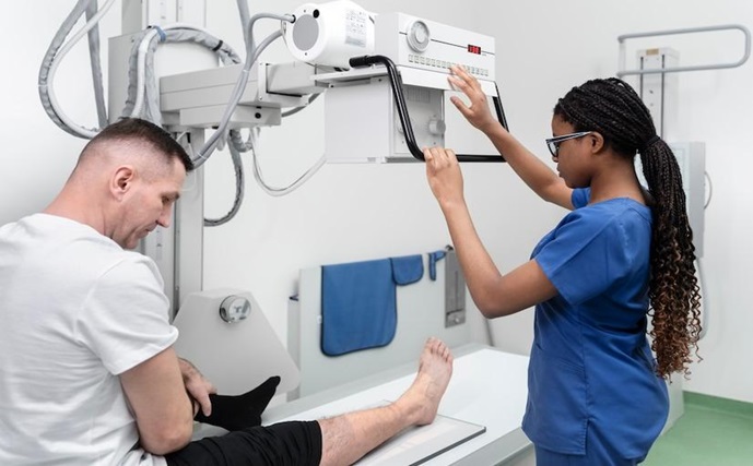
The Process Of Obtaining An MRI for Foot Diagnostics
If your doctor recommends an MRI for a foot injury, they will assess your symptoms and medical history first. If an MRI is needed, you’ll schedule it at an imaging center. Be sure to inform the staff of any implanted devices or medical concerns.
On the day of the MRI, you’ll change into a gown and remove any metal items. The scan takes 30 to 60 minutes, and you must remain still. The machine can be noisy, but earplugs or headphones will be provided. After the scan, you can resume normal activities unless advised otherwise.
Preparing For An MRI Scan
Follow your foot doctor’s or imaging center’s instructions to prepare for an MRI. Inform them of any medical conditions, allergies, or medications, particularly if you have kidney issues, as specific contrast agents may pose risks.
You may be asked to fast for a few hours before the scan, mainly if contrast is used. It’s usually safe to continue medications but confirm with your provider. For convenience, wear comfortable, metal-free clothing.
For those feeling anxious or claustrophobic, practice relaxation techniques like deep breathing or visualization, and consider bringing a supportive friend or family member.
What To Expect During An MRI Scan For Foot Diagnostics
During an MRI for foot diagnostics, you will lie on the table with your foot positioned inside the machine. You may need to keep your foot still for clear images. The machine will produce loud banging and thumping noises, which are expected, and you’ll be provided earplugs or headphones for comfort.
The scan lasts 30 to 60 minutes, during which you should remain still. Occasionally, you may be asked to hold your breath briefly. If you feel uncomfortable, you can communicate with the technician via an intercom, and they will monitor your comfort and safety throughout the procedure.
Interpreting MRI Results For Foot Fractures And Soft Tissue Damage
After the MRI scan, a radiologist will analyze the images and prepare a report for your foot doctor. The doctor will then discuss the results with you in a follow-up appointment.
The radiologist will identify fractures, their location, type, and severity, and check for any associated soft tissue injuries, such as ligament or tendon damage. This detailed analysis helps your doctor understand the full extent of the injury and plan the appropriate treatment.
The MRI may reveal inflammation, tears, or other ligaments, tendons, and cartilage issues for soft tissue damage, guiding treatment decisions, whether surgical, physical therapy or conservative management.
Treatment Options For Foot Fractures And Soft Tissue Damage
MRI results guide a tailored treatment plan for foot fractures and soft tissue damage. Minor injuries may require RICE, while severe fractures might need a cast, walking boot, or surgery to realign bones or repair tissues. Physical therapy and NSAIDs support recovery, aiming to restore function, prevent future injuries, and return to daily activities pain-free.
The Role Of Foot Doctors In Utilizing MRI for Diagnostics
Foot doctors are essential in MRI technology to diagnose and treat foot injuries. Their expertise in foot and ankle anatomy helps them interpret MRI results accurately, leading to precise diagnoses. After evaluating patients and reviewing MRI scans, they create personalized treatment plans, including rehabilitation exercises and lifestyle changes, to support recovery and ensure optimal outcomes.
Conclusion: The Future Of MRI In Foot Diagnostics
The future of MRI in foot diagnostics looks promising, with ongoing advancements in imaging techniques likely to enhance scan quality and precision. Innovations like functional MRI and improved contrast agents may offer deeper insights, enabling more accurate diagnoses and tailored treatments. Additionally, integrating artificial intelligence in interpreting MRI scans could lead to faster, more precise results, helping identify injuries that human radiologists might miss. As technology evolves, MRI will continue to be a vital tool for foot doctors, improving patient care and outcomes.
Comments are closed.